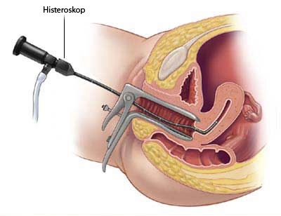
Hysteroscopy
Hysteroscopy is a method that enables to examine the intra uterine (uterine cavity) with the help of optical tools. In hysteroscopy, as in laparoscopy, an illuminated – optical system called a telescope is used. CO2 or special fluids are introduced from the cervix through a hysteroscope, so that one of the walls of the uterus is separated from the other. Direct imaging of the structures in the expanding uterus is provided by hysteroscopy.
Hysteroscopy can be used for diagnostic purposes (Diagnostic Hysteroscopy) or for treatment purposes (Operative Hysteroscopy).
1-Diagnostic Hysteroscopy; It is a procedure that can usually be performed without the need for anesthesia or without hospitalization with local anesthesia.

It is often called Office Hysteroscopy, and the tools used to enter the uterus are smaller than 5 mm and for easier evaluation of the inside of the uterus, it is mostly necessary to apply immediately after menstruation. Office hysteroscopy is an important tool in the investigation and treatment of the cause of infertility, recurrent miscarriage, and abnormal menstruation.
2-Operative Hysteroscopy; It is used for the many treatments such as Polyps detected in the uterus, fibroids, intrauterine adhesions, correction of uterine abnormalities (Septum) abnormal uterine bleeding, removal of foreign bodies in the uterus, etc. The diameter of the hysteroscope used here is 9 mm, and is designed to allow scissors, biopsy forceps, capture forceps, laser fibers and electrosurgical tools to be used in the operation to pass through the channels inside. Adhesions, fibroids and polyps that can be seen in the uterus can be removed. After surgery, a spiral (intrauterine device) or a thin urine probe (foley catheter) can be inserted into the uterus to prevent the walls of the uterus from sticking together, or specially prepared gels can be placed. Antibiotics and / or hormonal drugs can be used to prevent infection and accelerate the healing of the inner lining of the uterus.
